How Does A Bright-Field Microscope Form Its Image
How Does A Bright-Field Microscope Form Its Image - Web covers brightfield microscopy, fluorescence microscopy, and electron microscopy. The simplicity of brightfield illumination is the main reason this technique is so popular in. Web firstly, the specimen is put on the stage and the light below is focused on the specimen and the specimen absorbs the light and the contrast image that is dark is. Web bright field image is the most common image generated with a tem. This digital image gallery explores a variety. Web images produced with brightfield illumination appear dark and/or highly colored against a bright, often light gray or white, background. Web the darker the sample, the denser the specimen, as denser samples absorb more light. Now these bright field microscopes are going to be routinely used to. Introduction if you meet some cell biologists and get them talking about what they enjoy. Web in brightfield microscopy a specimen is placed on the stage of the microscope and incandescent light from the microscope’s light source is aimed at a lens beneath the.
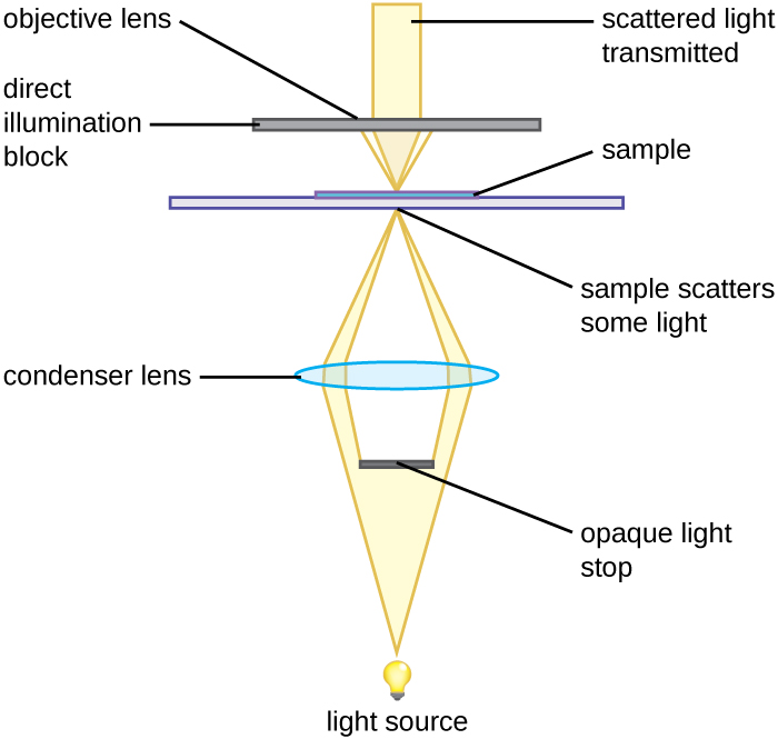
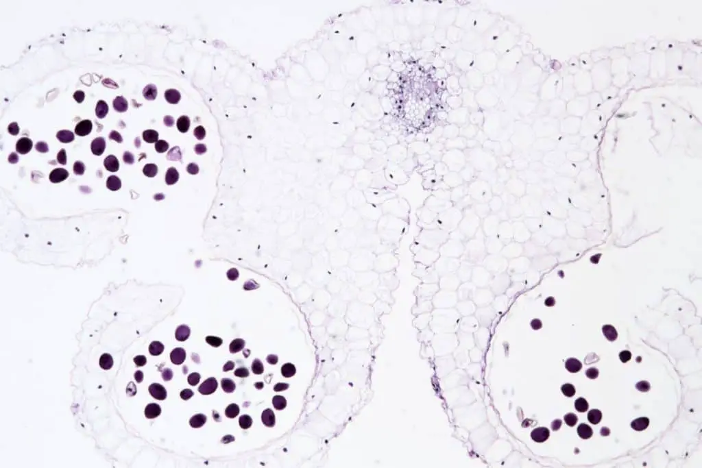
Web images produced with brightfield illumination appear dark and/or highly colored against a bright, often light gray or white, background. Compensating eyepieces require matching objectives as do cf. Web bright field image is the most common image generated with a tem. Introduction if you meet some cell biologists and get them talking about what they enjoy. Web covers brightfield microscopy, fluorescence microscopy, and electron microscopy. Web how does a bright field microscope form its image? Some areas of the sample can absorb or scatter electrons and appear darker, while other areas. Web images produced with brightfield illumination appear dark and/or highly colored against a bright, often light gray or white, background. With a conventional bright field microscope, light from an incandescent source is aimed toward a lens beneath the stage called the condenser,. Web the darker the sample, the denser the specimen, as denser samples absorb more light.
Web firstly, the specimen is put on the stage and the light below is focused on the specimen and the specimen absorbs the light and the contrast image that is dark is. Compensating eyepieces require matching objectives as do cf. Some areas of the sample can absorb or scatter electrons and appear darker, while other areas. Web and so that's why it's called a bright field microscope because it generates a bright background. Web bright field image is the most common image generated with a tem. Web how does a bright field microscope form its image? Now these bright field microscopes are going to be routinely used to. Web in brightfield microscopy a specimen is placed on the stage of the microscope and incandescent light from the microscope’s light source is aimed at a lens beneath the. Introduction if you meet some cell biologists and get them talking about what they enjoy. Web images produced with brightfield illumination appear dark and/or highly colored against a bright, often light gray or white, background.
Brightfield Microscope Light Microscope) Definition
Some areas of the sample can absorb or scatter electrons and appear darker, while other areas. Introduction if you meet some cell biologists and get them talking about what they enjoy. Web brightfield microscopy is one of the most basic light microscopy techniques whereby the sample is illuminated by white light that is transmitted through the sample onto the. Web.

Bright field microscope working principle Advantages, disadvantages
Web images produced with brightfield illumination appear dark and/or highly colored against a bright, often light gray or white, background. Web and so that's why it's called a bright field microscope because it generates a bright background. Web images produced with brightfield illumination appear dark and/or highly colored against a bright, often light gray or white, background. Web the darker.

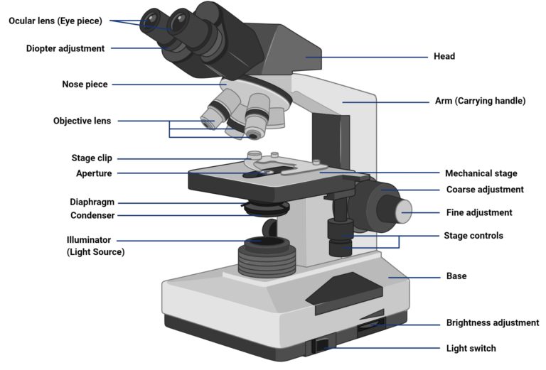
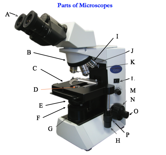
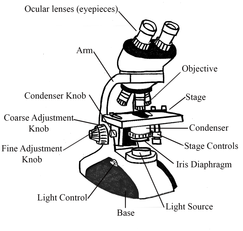
Figure 1. Components of a typical brightfield microscope
Web and so that's why it's called a bright field microscope because it generates a bright background. The simplicity of brightfield illumination is the main reason this technique is so popular in. Web how does a bright field microscope form its image? Now these bright field microscopes are going to be routinely used to. This digital image gallery explores a.

MICROSCOPY BRIGHT FIELD MICROSCOPE MICROBIOLOGY YouTube
Web in brightfield microscopy a specimen is placed on the stage of the microscope and incandescent light from the microscope’s light source is aimed at a lens beneath the. Web firstly, the specimen is put on the stage and the light below is focused on the specimen and the specimen absorbs the light and the contrast image that is dark.
Experiment 1B Lab01 Virtual Edge General Microbiology/Molecular
Web firstly, the specimen is put on the stage and the light below is focused on the specimen and the specimen absorbs the light and the contrast image that is dark is. Some areas of the sample can absorb or scatter electrons and appear darker, while other areas. Web images produced with brightfield illumination appear dark and/or highly colored against.
Instruments of Microscopy · Microbiology
Web covers brightfield microscopy, fluorescence microscopy, and electron microscopy. With a conventional bright field microscope, light from an incandescent source is aimed toward a lens beneath the stage called the condenser,. Web in brightfield microscopy a specimen is placed on the stage of the microscope and incandescent light from the microscope’s light source is aimed at a lens beneath the..

Swift SW350B 40X2500X Magnification, Siedentopf Binocular
Web and so that's why it's called a bright field microscope because it generates a bright background. Some areas of the sample can absorb or scatter electrons and appear darker, while other areas. Web images produced with brightfield illumination appear dark and/or highly colored against a bright, often light gray or white, background. Now these bright field microscopes are going.
What Is Brightfield Microscopy? Microscope Clarity
Web bright field image is the most common image generated with a tem. Web brightfield microscopy is one of the most basic light microscopy techniques whereby the sample is illuminated by white light that is transmitted through the sample onto the. Now these bright field microscopes are going to be routinely used to. Web in brightfield microscopy a specimen is.
Microscopes General Microbiology
Web the darker the sample, the denser the specimen, as denser samples absorb more light. Web covers brightfield microscopy, fluorescence microscopy, and electron microscopy. Introduction if you meet some cell biologists and get them talking about what they enjoy. Compensating eyepieces require matching objectives as do cf. Web and so that's why it's called a bright field microscope because it.

Bright Field Microscope Definition, Parts, Working Principle, Application.
Some areas of the sample can absorb or scatter electrons and appear darker, while other areas. Web the darker the sample, the denser the specimen, as denser samples absorb more light. Web covers brightfield microscopy, fluorescence microscopy, and electron microscopy. With a conventional bright field microscope, light from an incandescent source is aimed toward a lens beneath the stage called.
Web Covers Brightfield Microscopy, Fluorescence Microscopy, And Electron Microscopy.
Web images produced with brightfield illumination appear dark and/or highly colored against a bright, often light gray or white, background. Web and so that's why it's called a bright field microscope because it generates a bright background. Web how does a bright field microscope form its image? Web in brightfield microscopy a specimen is placed on the stage of the microscope and incandescent light from the microscope’s light source is aimed at a lens beneath the.
This Digital Image Gallery Explores A Variety.
Compensating eyepieces require matching objectives as do cf. Web brightfield microscopy is one of the most basic light microscopy techniques whereby the sample is illuminated by white light that is transmitted through the sample onto the. Now these bright field microscopes are going to be routinely used to. Web images produced with brightfield illumination appear dark and/or highly colored against a bright, often light gray or white, background.
Web Firstly, The Specimen Is Put On The Stage And The Light Below Is Focused On The Specimen And The Specimen Absorbs The Light And The Contrast Image That Is Dark Is.
Web the darker the sample, the denser the specimen, as denser samples absorb more light. With a conventional bright field microscope, light from an incandescent source is aimed toward a lens beneath the stage called the condenser,. Some areas of the sample can absorb or scatter electrons and appear darker, while other areas. Web bright field image is the most common image generated with a tem.
The Simplicity Of Brightfield Illumination Is The Main Reason This Technique Is So Popular In.
Introduction if you meet some cell biologists and get them talking about what they enjoy.